An aesthetically pleasing eye has an almond shape with superior arc that peaks medially 27 and a slight upward inclination of the lateral canthal angle positive canthal tilt. They argue that canthal anchoring sutures are not needed permanently and using absorbable sutures prevents the risk of stitch sinus or infection.
Revisional Eyelid Surgery Fixing The Canthus Dr Guy Massry
Fronto-ethmoidal external approaches and more rarely external DCR and blepharoplasty represent the commonest iatrogenic causes of medial canthal webbing.

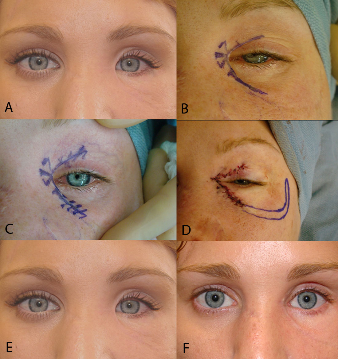
. Medial canthal web after upper blepharoplasty. Determine the position of the lower eyelid and lateral canthus after release of the lower lid retractors with the inside-out technique by measuring the MR2 and use of the lateral canthal rounding scale. The scars usually occur when the incisions are carried too medially and the skin bridges the supero-medial hollow of the upper lid in a straight line.
Webs abnormal folds of skin can occur in both areas and are referred to as medial and lateral canthal webs. Do it at least 3 times a day with 3 repeatitions and 20-30 seconds hold in each more Sorry about your problem. Canthal webbing can be associated with scleral show laterally due to inferior lid retraction and is a known complication of blepharoplasty or reconstruction following trauma or tumour excision 1.
This area near the nose is called the medial canthus and the same area on the outer eyelids is called the lateral canthus. I recommend Z-plasty repair of the medial canthal webbing. WARNING Some photos may be explicit.
Unrecognized or untreated lower lid laxity may contribute to well-recognized deformities after aesthetic eyelid surgery such as the round-eye syndrome canthal malposition and scleral show. Generally for patients having almond eyes with a thick medial canthus folds at one-fifth of the inner canthus are naturally embedded in the. The scars usually occur when the incisions are carried too medially and the skin bridges the supero-medial hollow of the upper lid in a straight line.
Medial canthus Mohs defect. After glabellar flap repair. A thorough understanding of the upper eyelid anatomy is essential when evaluating patients for possible upper blepharoplasty.
Secondary lateral canthal webs may occur after trauma or blepharoplasty. For an upper lid blepharoplasty ending the incision just lateral to the punctum avoids medial canthal webbing as well as lacrimal system injury. It provides excellent cosmesis and is associated with minimal complications.
The cosmetic result was highly satisfactory in all cases. An effective preventive measure is to taper the nasal incision markings superiorly and to avoid extending the marking medially to the punctum. May be due to incision extended too far medially.
Therefore many patients choose to undergo concurrent medial canthoplasty and double eyelid surgery to maximize the postoperative results. Every patient voiced aesthetic concerns with the web and 4 57 of the 7 patients with lateral canthal webs. Canthal webs occur because there is a relative deficiency of skin in the vertical as compared to the horizontal plane.
It is very had to tell from your photos if you have any canthal webbing. Everyone has seen their eyes be tired and puffy but some people have to live with that. Four procedures are described to repair these webs.
Postoperative canthal webbing is aesthetically displeasing and may be noticed by the patient andor surgeon. Besides enlarging the eyes medial canthoplasty changes the eye shape or double eyelid patterns. Seven 88 of the patients had lateral canthal webs after surgery and 1 12 patient had a medial canthal web after a motor vehicle accident.
If it persists then revision by Y-Vtype incisions is warranted. Download high-res image 98KB Download. Age-related attenuation of the canthal constituents particularly the tarsoligamentous imparts laxity to the lower eyelid.
The best way to handle it is to do a push-pull massage of the surgical area. There were no major complications or re-operations. It does look like you have some sclera like show on the right and scarring of the lower lid lateral canthus on the left.
And blepharoplasty represent the commonest iatrogenic causes of medial canthal webbing. The skin and orbicularis oculi muscle form the anterior layers of the upper eyelid. All clinical photographs are actual patients of Dr.
Do I have any good options. Ad When it comes to blepharoplasty or surgery of the eyelids there are certain things that. Large basal cell carcinoma.
Massage and steroid injections can help. It requires medial canthal scar revision with multiple z-plasty. Less Sorry about your problem.
Two cases had minor webbing of the medial upper lid. Large basal cell carcinoma. They are surgically corrected by a variety of micro-skin flaps.
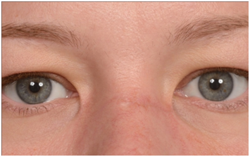
The palpebral fissure shape and dimensions should be preserved and sometimes corrected during blepharoplasty. Canthal web revision Canthoplasty Revision Canthoplasty The area where the upper and lower lids meet is called the canthus. Medial canthus Mohs defect.
It forms a c shape and makes my eyes asymmetrical. In cases of canthoplasty the upper and lower eyelid should be carefully and precisely aligned with 6-0 buried vicryl sutures to prevent canthal webbing. I have inner eyelid webbing following a blepharoplasty 2 years ago.
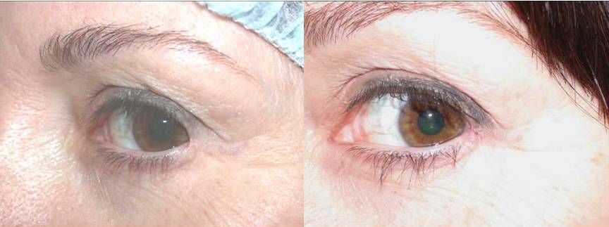
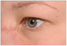
Incisions should be at least 45 mm above the punctum to avoid the canaliculus. The angle and shaddowing make it difficult to determine exactly what is going on. Download scientific diagram Medial canthal webbing seen after upper lid blepharoplasy done by a dermatologist.
May be corrected by Zplasty Wplasty transposition flaps or YV advancement procedures. May be due to inadvertent trauma to the levator complex including postsurgical edema and dehiscence. If a medial canthal web does result time massage and steroid injections can help.
Medial canthal webbing can be revised with a Z-plasty. The punctum is a useful landmark for the upper lid and lower lid incision. 3 The lateral canthal angle is sharp and crisp with the lateral commissure closely opposed.
Anatomy of the upper eyelid. Most plastic surgeons avoid addressing canthal webs as they are difficult to. The rhomboid flap is an effective quick and simple technique for medial canthal reconstruction.
Please see beforeafter photo on link below toward bottom of the website page. Deep to these layers is the orbital septum which originates from the arcus marginalis at the superior. After glabellar flap repair.
Prevent by planning an incision that extends to the medial commissure. Post-blepahroplasty webbing can be seen when upper and lower blepharoplasty is performed together and the lateral skin incisions of the procedures are in close proximity less than 5 mm apart. In some cases early recognition and aggressive massage can result in better aesthetic results by improving.
78 of the 171 patients with the inside out blepharoplasty had follow up of 3 months. However these complications are uncommon.
![]()
Medial Canthal Webbing Seen After Upper Lid Blepharoplasy Done By A Download Scientific Diagram

Medial Canthal Webbing Seen After Upper Lid Blepharoplasy Done By A Download Scientific Diagram
Case Of Bilateral Iatrogenic Medial Canthal Webbing Treated With Full Thickness Skin Grafts Medcrave Online

Medial Canthal Webbing Seen After Upper Lid Blepharoplasy Done By A Download Scientific Diagram
Medial Canthal Webbing Seen After Upper Lid Blepharoplasy Done By A Download Scientific Diagram

Medial Canthal Webbing Seen After Upper Lid Blepharoplasy Done By A Download Scientific Diagram
Case Of Bilateral Iatrogenic Medial Canthal Webbing Treated With Full Thickness Skin Grafts Medcrave Online
Canthal Web Revision Canthoplasty Revision Canthoplasty Dr Guy Massry
0 comments
Post a Comment